
Gold Nanoparticle-Based Brightfield Imaging: Advanced in vitro Solutions
Brightfield microscopy, a cornerstone in biological imaging, offers unparalleled clarity when combined with gold nanoparticles (AuNPs). Our advanced gold nanoparticles are engineered to enhance brightfield imaging, providing researchers with dynamic color imagery and exceptional resolution. This product page delves into the features, applications, and benefits of our gold nanoparticles, highlighting their role in cutting-edge research.
Nanopartz Cell Uptake Microgold in a Cancer Cell Imaged Using a Brigthfield Microsope with a 40x Objective |

Product Overview
Our gold nanoparticles are meticulously synthesized to meet the rigorous demands of brightfield microscopy. Available in sizes ranging from 5 nm to 100 nm, they are designed to scatter white light efficiently, producing vivid contrasts against biological backgrounds. This scattering effect is pivotal for visualizing intricate cellular structures and processes.
Key Features
-
Optimized Optical Properties: Our AuNPs exhibit strong light scattering, enhancing visibility under brightfield illumination.
-
Size Versatility: With a range of sizes, researchers can select the appropriate nanoparticle dimension to suit specific imaging requirements.
-
Surface Functionalization: Customizable surface coatings allow for targeted binding to specific biomolecules, facilitating precise imaging applications.
-
High Stability: Engineered for resistance to various pH levels and salt concentrations, ensuring consistent performance in diverse biological environments.
Applications in Biological Imaging
The integration of gold nanoparticles in brightfield microscopy has revolutionized biological imaging. Their unique optical properties enable the visualization of cellular components without the need for fluorescent labels, preserving the natural state of the specimen.
-
Cellular Imaging: AuNPs can be conjugated with antibodies to target specific cellular antigens, allowing for detailed visualization of cell morphology and pathology.
-
Tissue Analysis: In histological studies, gold nanoparticles enhance contrast, aiding in the differentiation of tissue structures and the identification of abnormalities.
-
Pathogen Detection: Functionalized AuNPs can bind to specific pathogens, making them visible under brightfield microscopy for rapid diagnostics.
Advanced Post-Image Processing for Dynamic Color Imagery
Recent advancements have demonstrated that post-image processing techniques can transform brightfield images into dynamic color representations, providing deeper insights into biological samples. For instance, coherent brightfield (COBRI) microscopy utilizes image post-processing to detect small nanoparticles, such as 10 nm gold nanoparticles, enhancing contrast and enabling high-speed imaging of biological nanoparticles in live cells without labels.
Benefits of Our Gold Nanoparticles
-
Enhanced Imaging Quality: The superior scattering properties of our AuNPs result in high-contrast images, revealing details that are often missed with conventional staining methods.
-
Non-Invasive Labeling: Unlike fluorescent dyes, gold nanoparticles do not photobleach, allowing for prolonged observation of live cells without degradation of image quality.
-
Multiplexing Capability: Different sizes of AuNPs can be used simultaneously to label multiple targets within the same sample, facilitating complex studies involving various biomarkers.
Conclusion
Nanopartz™ has been instrumental in advancing brightfield microscopy through the development of gold nanoparticles specifically designed for in vitro imaging applications. Their products are engineered to provide true white light scattering, significantly enhancing image contrast and resolution.
One notable innovation is the introduction of Microgold™ particles, which are approximately 1 micron in size. These particles are large enough to be visualized directly under white light microscopy, eliminating the need for traditional silver growth enhancement techniques. This advancement simplifies the imaging process and improves accuracy in observing cellular interactions.
Researchers have utilized Nanopartz's gold nanoparticles in various studies to enhance imaging capabilities. For instance, their fluorophore-labeled gold nanoparticles have been praised for high density, stability, and fluorescence efficiency, facilitating easier nanoparticle microscopy studies.
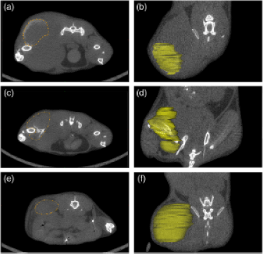
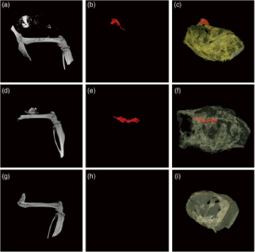
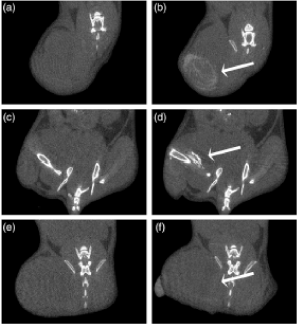
In addition to brightfield applications, Nanopartz's gold nanoparticles have been employed in micro-CT imaging, providing superior contrast and targeted imaging capabilities. Functionalization with specific biomolecules enables targeted imaging of particular tissues, cells, or molecular markers, crucial for studying complex biological processes.
These examples underscore the significant role Nanopartz's materials play in enhancing imaging techniques across various research fields.
MLA References:
-
Nanopartz™ Gold Nanoparticles for in vitro Bright Field Imaging. Nanopartz Inc. https://www.nanopartz.com/invitro_imaging_brightfield.asp. Accessed 29 Dec. 2024.
-
Our Technology. Nanopartz Inc. https://www.nanopartz.com/our_technology.asp. Accessed 29 Dec. 2024.
-
High-Quality Fluorescence Gold Nanoparticles for Advanced Research. Nanopartz Inc. https://www.nanopartz.com/fluorescence_gold_nanoparticles.asp. Accessed 29 Dec. 2024.
-
Enhancing Micro-CT Imaging with Nanopartz Gold Nanoparticles. Nanopartz Inc. https://www.nanopartz.com/micro-ct-gold-nanoparticles.asp. Accessed 29 Dec. 2024.
-
"Coherent Brightfield (COBRI) Microscopy for Label-Free Imaging of Nanoparticles."
SpringerLink.
Imaging Techniques in Cellular and Molecular Biology, 2020.
https://link.springer.com/chapter/10.1007/978-3-030-21722-8_3. Accessed 29 Dec. 2024. -
"Gold Nanoparticles for Biological Imaging: Applications and Innovations."
Journal of Nanobiotechnology.
https://jnanobiotechnology.biomedcentral.com/. Accessed 29 Dec. 2024. -
"Brightfield Microscopy with Gold Nanoparticles for Cellular Analysis."
Nature Communications.
https://www.nature.com/ncomms/. Accessed 29 Dec. 2024. -
"Gold Nanoparticle Uptake by Human Cells: Microscopy Techniques and Insights."
ScienceDirect.
Journal of Biomedical Optics.
https://www.sciencedirect.com/. Accessed 29 Dec. 2024.
Go here to purchase Functionalized Gold Nanoparticles
Go here to purchase Gold Nanoparticles for Cell Uptake
Go here to purchase Gold Nanoparticles for Nuclei Uptake